Background
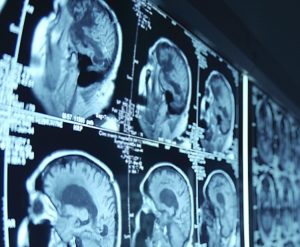
The gold standard for examining hepatocellular carcinoma (HCC) is biopsy, which is an invasive procedure that can often result in morbidity/mortality. In addition, the locations for biopsy samples are limited within the liver or a mass and thus suffer from sampling errors. While multiple imaging modalities have been used, liver disease diagnosis has gradually transitioned from ultrasonography (US) and computed tomography (CT) to magnetic resonance imaging (MRI). However, a retrospective analysis found that imaging-based diagnosis tends to result in a high number of false positives labeling common benign focal abnormalities in diseased liver as HCC.
Objective
This project aims to fully investigate an innovative technique, MRE coupled with novel computational tools, for early and reliable HCC diagnosis. The objective is to show that MRE can accurately:
-
determine the risk of developing HCC; including identifying independently associated factors (e.g., inflammation and fibrous tissue)
-
discriminate HCC from nonmalignant cirrhotic nodules (e.g., regenerative and dysplastic) and mimics (e.g., perfusion anomalies)
-
assess response to therapy at timepoints earlier than conventional imaging techniques
-
predict risk of HCC recurrence
Approach
Innovative computational tools will be developed to extract imaging signatures and create a robust machine learning model. This model will optimally utilize the information available from MRE (both kPa and imaging signatures) to study HCC behavior. To validate the results in clinical usage, the multi-parametric approach with MRE will be correlated with histology (biopsy/resection, explant), traditional tumor response assessment algorithms (RECIST, mRECIST), and specific clinical end-points (e.g., time-to-disease-progression and disease survival).
Impact
MRE has not been robustly tested in clinical applications for HCC, and its implementation has been limited mostly to academic centers, such as Mayo Clinic. Thus, there are a relatively small number of studies in the literature investigating the efficacy of this novel technique for HCC; most of which are based on phantom and animal models. Though MRE has been explored in evaluating hepatic fibrosis in smaller in vivo studies, the studies have mainly focused on diffuse liver disease instead of HCC.
HCC prognosis remains poor due to suboptimal characterization and therapeutic response assessments with current imaging techniques that rely on morphologic and differential dynamic enhancement changes. The proposed approach will utilize MRE techniques that can evaluate extracellular matrix biochemical composition for evaluating, and potentially modifying, HCC behavior. Most research on MRE focuses on the fibrosis score (kPa), a measure of mechanical properties of tissue. It is believed that image intensity describing the local characteristic patterns can be of help in identifying tissue composition. This image intensity may also be useful in HCC diagnosis.
Publications
-
Silva, A., Grimm R., Glaser K., *Fu, Y., Wu, T., Ehman R., Silva, C., “Magnetic Resonance Elastography: Evaluation of New Inversion Algorithm and Quantitative Analysis Method”, Abdominal Imaging, April, 2015, 40 (4): 810-7. doi: 10.1007/s00261-015-0372-5.