Cancer is the leading cause of death of Americans aged 40-79 years. Part of the problem is ineffective evaluation of treatment responses. Currently applied tumor response metrics, such as Response Evaluation Criteria in Solid Tumors, rely on subjective tumor measurements with high intra- and inter- observer variability, necessitating a wide margin of change for response classification. The goal of this project is to demonstrate that image-based measurement of therapy response can be improved by standardizing procedures and using advanced imaging analytic tools.
Methods
The proposed study was conducted in the iCORE (imaging Clinical Outcomes and Response Evaluation) laboratory housed in the department of radiology, MCA. We completed two interconnected tasks:
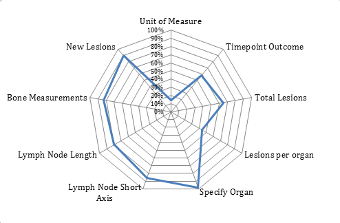
Task I: Evaluate clinical (non-iCORE) vs. iCORE reports generated using Mint 3.0 (Mint Medical, Dossenheim, Germany) to assess impact on clinical trial decision making. Conformity error; inter- & intra-observer variability; measurement time duration and slice level consistency were evaluated.
Task II: Exploring advantages of quantitative imaging (QI) tools in improving diagnosis. Diagnostic accuracy, sensitivity and specificity were evaluated.

Source: ASU-Mayo Science of Healthcare Delivery